Hidden angina check which can identify early warning signs
Date Posted: 16th December 2019

Highly specialist scan shows whether arteries supplying blood to the heart are narrowed or becoming blocked.
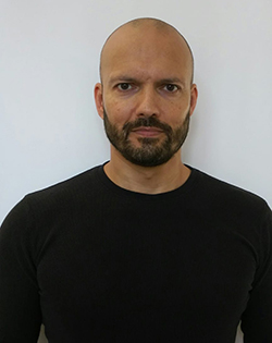
We spoke to Chris, a patient at St Joseph’s who recently had a CT Coronary Angiogram, to ask him a few questions about why he chose to have his heart assessed. We then spoke to the experts at St Joseph's to find out what the CT scan identified.
Tell us a little about your lifestyle and how you like to keep fit and active?
I enjoy an active lifestyle walking, cycling and playing golf when time allows. I’ve cycled in charity bike rides for Children’s Hospice South West and plan to cycle from Land’s End to John o’ Groats in June 2020 which is one of the reasons why I decided to have a heart check.
Does your family have a history of heart problems?
Yes, my grandfather had his first heart attack at 58 followed by a fatal heart attack at the age of 63. My uncle had triple heart bypass surgery at 60 and my father had heart surgery in his late 70’s.
What made you choose St Joseph’s Hospital for a CT scan?
I did some research into the level of technology available for heart scanning. St Joseph’s Hospital has the 320-Slice CT Scanner which amazingly images the entire heart in a single heartbeat. It also gives the lowest radiation dose possible when it comes to CT scanning. The scan looks at the arteries which supply the blood to the heart and identifies any narrowing or potential blockages. I also wanted to know that I’d be looked after by a specialist and was impressed by the level of expertise available at St Joseph’s.
What questions were you asked before the scan by the specialists?
I had to fill in a full health questionnaire and be assessed by a specialist. My family history of heart disease was a significant factor when deciding on the CT scan.
Were you nervous at all?
I didn’t have any concerns about the procedure but I was a little nervous about the outcome.
How were you treated before and after the procedure?
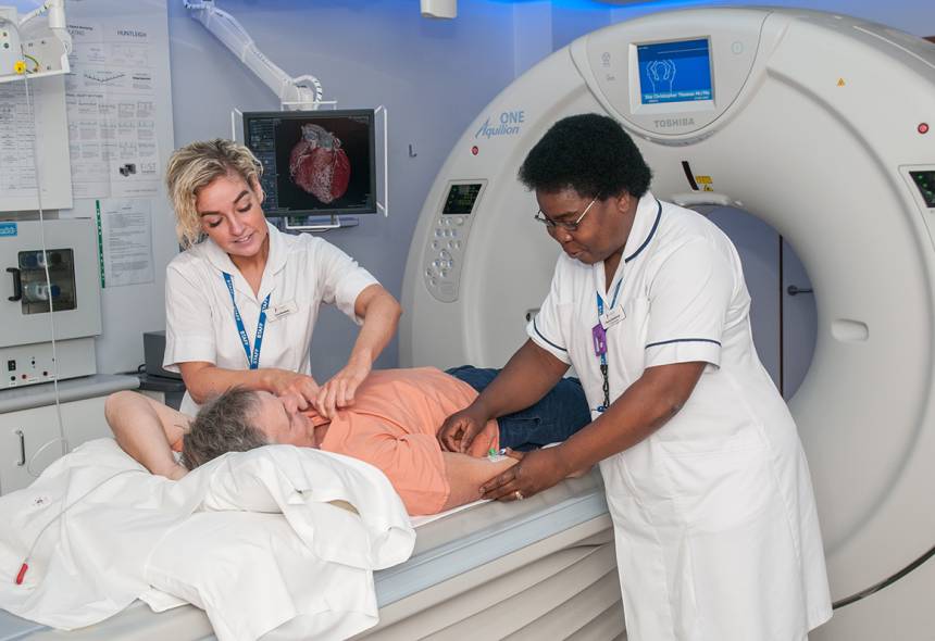
The Radiographers, Charity and Lucy, were amazing. They were highly professional but with the perfect level of humour to match – they made me feel at ease and relaxed.

Is it strange seeing a detailed 3D image of your heart?
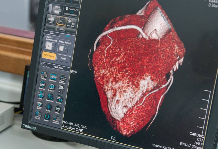
Yes very strange indeed. Before the scan, I was given medicine to slow my heart rate, but you stay awake. A small needle was inserted into my arm and used to administer the X-ray dye during the procedure. I had to lie on a bed which moved through the scanner.
Was everything ok with your scan?
My scan was not totally normal but nothing to cause any immediate action.
It identified ‘early’ furring of the arteries and mild aortic root dilatation with a recommendation to have my cholesterol levels checked and an MRI scan of my aorta in 12 months’ time. At least I now know, at the of 52, when I have time to do something about my cholesterol levels.
Would you recommend this service to others?
We all bury our heads in the sand, especially men. If you’re given the chance to change your life for the better why would you not do it? Most importantly you get to meet Charity who will fill you with life; her passion for what she does is extraordinary. The hospital offers a very warm and professional service.
Expert opinion; the results explained

Dr Nathan E. Manghat, Consultant Cardiovascular and Interventional Vascular Radiologist explains; The CT scan for Chris detected the presence of coronary artery disease in the vessel walls. This precedes a significant narrowing which then reduces the blood supply to the heart muscle.
"We also know that the shallow, more fatty arterial disease is more likely to present with a heart attack as the first symptom. The detection of early disease in patients at increased risk can ensure that the correct preventative medication is started with advice on appropriate lifestyle modification.
“It also identified Atheroma or ‘furring of the arteries’. This is a complex process caused by inflammatory blood cells and fatty/cholesterol build-up within the arterial walls; the end result of progressive inflammation and re-healing is the production of harder calcium deposits in the blood vessels which can also contribute to narrowing the calibre of blood vessels or sometimes cause them to dilate and form ‘aneurysms’ – this process can occur in all arteries of the body which can affect the blood supply to all organ systems.
“The incidental detection of mild aortic root dilatation was important; the major blood vessel leaving the heart can get bigger for the same reasons that we get furring up of the arteries but it can also be due to heart valve disease and can be hereditary. If the blood vessel gets bigger, the risk of
the wall tearing (dissection), or rupture increases. This can lead to sudden death if not detected or followed up appropriately.
“Whilst we cannot predict the future, the high-end MRI and CT scanner technology available at St Joseph’s Hospital helps us to provide the most accurate imaging data possible to assist in the diagnosis of potentially high risk cardiovascular disease. Our cardiac analysis not only may provide the welcome reassurance that our patients are seeking, but may also facilitate best medical therapy, advice and appropriate clinical follow-up in order to reduce future risk.”


